Beranda
/ Anatomy Of The Upper Chest Area - Chest Anatomy Artwork Stock Photo Alamy / The point of origin of chest pain can be any one of the organs in the chest, namely heart, lung, or esophagus, or from the components of the chest wall.
Anatomy Of The Upper Chest Area - Chest Anatomy Artwork Stock Photo Alamy / The point of origin of chest pain can be any one of the organs in the chest, namely heart, lung, or esophagus, or from the components of the chest wall.
Insurance Gas/Electricity Loans Mortgage Attorney Lawyer Donate Conference Call Degree Credit Treatment Software Classes Recovery Trading Rehab Hosting Transfer Cord Blood Claim compensation mesothelioma mesothelioma attorney Houston car accident lawyer moreno valley can you sue a doctor for wrong diagnosis doctorate in security top online doctoral programs in business educational leadership doctoral programs online car accident doctor atlanta car accident doctor atlanta accident attorney rancho Cucamonga truck accident attorney san Antonio ONLINE BUSINESS DEGREE PROGRAMS ACCREDITED online accredited psychology degree masters degree in human resources online public administration masters degree online bitcoin merchant account bitcoin merchant services compare car insurance auto insurance troy mi seo explanation digital marketing degree floridaseo company fitness showrooms stamfordct how to work more efficiently seowordpress tips meaning of seo what is an seo what does an seo do what seo stands for best seotips google seo advice seo steps, The secure cloud-based platform for smart service delivery. Safelink is used by legal, professional and financial services to protect sensitive information, accelerate business processes and increase productivity. Use Safelink to collaborate securely with clients, colleagues and external parties. Safelink has a menu of workspace types with advanced features for dispute resolution, running deals and customised client portal creation. All data is encrypted (at rest and in transit and you retain your own encryption keys. Our titan security framework ensures your data is secure and you even have the option to choose your own data location from Channel Islands, London (UK), Dublin (EU), Australia.
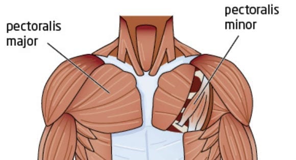
Anatomy Of The Upper Chest Area - Chest Anatomy Artwork Stock Photo Alamy / The point of origin of chest pain can be any one of the organs in the chest, namely heart, lung, or esophagus, or from the components of the chest wall.. The dominant muscle in the upper chest is the pectoralis major. Iv contrast may be injected into a vein in the patient's arm or hand. The pec major) is the one that commands the most real estate. Related posts of anatomy of the chest area. In other words, each area does something different.
Root of lung , superior lobe; The spinal cord represents the cns in the thorax and serves as the vital link between the brain and the body. The 3 top exercises to build the upper chest 3. Of the two chest muscles, the pectoralis major (a.k.a. Chest pain can be divided into two types, namely right side chest pain and left side chest pain.
Abdomen Wikipedia from upload.wikimedia.org Iv contrast may be injected into a vein in the patient's arm or hand. This thoracic and pulmonary anatomy tool is especially designed for students of anatomy (medical and paramedical studies). The pectoralis minor (which is of little concern to us for now), the clavicular head of the pectoralis major. Dermatomes of the upper limbs are innervated by spinal nerves c5. The upper chest has two main functions: We're looking at the anatomy of an upper endoscopy. Anatomy of the chest and the lungs: 8 best upper chest exercises.
Related posts of anatomy of the chest area.
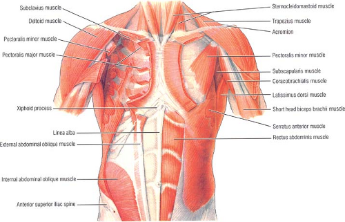
The chest is the area of origin for many of the body's systems as it houses organs such as the heart, esophagus, trachea, lungs, and thoracic diaphragm. 8 best upper chest exercises. The circulatory system does most of its. Browse 2,531 female chest anatomy stock photos and images available, or start a new search to explore more stock photos and images. The epidermis is the outermost layer that provides a protective, waterproof seal over the body. the division into the separate, distinct parts of this muscle is about functionality. Understanding the basics of throat anatomy with diagram and pictures. For the purpose of description the lungs are divided into zones: However, it is important to remember, that not every chest pain means a heart attack. This is the lowermost portion of the pecs. The muscles of the chest and upper back occupy the thoracic region of the body inferior to the neck and superior to the abdominal region and include the muscles of the shoulders. The chest is the area of origin for many of the body's systems as it houses organs such as the heart, esophagus, trachea, lungs, and thoracic diaphragm. The major muscle in the chest is the pectoralis major.
Chest pain can be divided into two types, namely right side chest pain and left side chest pain. 8 best upper chest exercises. Upper back pain and chest pain can occur together. I lost my job and my medical coverage and have been taking prednisone, 15mg per day, as a stop gap. The point of origin of chest pain can be any one of the organs in the chest, namely heart, lung, or esophagus, or from the components of the chest wall.
Applied Anatomy Of The Chest Wall And Mediastinum Basicmedical Key from basicmedicalkey.com Male internal anatomy of chest and abdominal area on black background. The clavicles are visible and palpable bony. The chest is the area of origin for many of the body's systems as it houses organs such as the heart, esophagus, trachea, lungs, and thoracic diaphragm. System respiratory respiratory organs of human body digestive and respiratory system medical chest internal structure of human body medicine body lungs biology intestines stomach anatomy torso human internal. Human internal organs dummy, training dummy, detail of the face, thorax and intestines. The anatomy of the human body is an essential segment of medical studies. The chest is part of a larger group of pushing muscles found in the upper body. Upper back pain and chest pain can occur together.
While it is only around one half of an inch (1 cm) in diameter, the spinal cord both carries nervous signals and processes many reflexes to support the structures of the body.
The upper fibers, the middle fibers (called the middle trapezius), and the lower fibers (called the lower traps). Understanding the basics of throat anatomy with diagram and pictures. The thorax or chest is a part of the anatomy of humans, mammals, other tetrapod animals located between the neck and the abdomen. For the purpose of description the lungs are divided into zones: The major muscle in the chest is the pectoralis major. The clavicles are visible and palpable bony. The 3 top exercises to build the upper chest 3. Chest pain can be divided into two types, namely right side chest pain and left side chest pain. To fully develop your chest, you need to hit it with heavy weight using a couple smartly chosen exercises. These important muscles control many motions that involve moving the arms and head — such as throwing a ball, looking up at the sky, and raising your hand. The upper chest has two main functions: The pectoralis minor (which is of little concern to us for now), the clavicular head of the pectoralis major. Male internal anatomy of chest and abdominal area on black background.
Browse 2,531 female chest anatomy stock photos and images available, or start a new search to explore more stock photos and images. Related posts of anatomy of the chest area. The pectoral region is located on the anterior chest wall. It contains four muscles that exert a force on the upper limb: The human thorax includes the thoracic cavity and the thoracic wall.
Chest Workout 5 Exercises To Build The Upper Chest Myprotein from blogscdn.thehut.net Tumour can be seen in the left upper lobe of his lungs man having defibrillation. For the purpose of description the lungs are divided into zones: Of the two chest muscles, the pectoralis major (a.k.a. The pec major) is the one that commands the most real estate. This thoracic and pulmonary anatomy tool is especially designed for students of anatomy (medical and paramedical studies). The pectoralis major, pectoralis minor, serratus anterior and subclavius. The muscles of the chest and upper back occupy the thoracic region of the body inferior to the neck and superior to the abdominal region and include the muscles of the shoulders. The clavicles are visible and palpable bony.
The spinal cord represents the cns in the thorax and serves as the vital link between the brain and the body.
As mentioned above, the trapezius muscle is divided into 3 areas: the division into the separate, distinct parts of this muscle is about functionality. The epidermis is the outermost layer that provides a protective, waterproof seal over the body. System respiratory respiratory organs of human body digestive and respiratory system medical chest internal structure of human body medicine body lungs biology intestines stomach anatomy torso human internal. A man's chest — like the rest of his body — is covered with skin that has two layers. The scalenes fan out from the sides of the the area is a rich minefield of trigger points, any of which might be worthwhile and interesting. In other words, each area does something different. In insects, crustaceans, and the extinct trilobites, the thorax is one of the three main divisions of the creature's body, each of which is in turn composed of multiple segments. To fully develop your chest, you need to hit it with heavy weight using a couple smartly chosen exercises. The throat is one of the most complex parts of the human body. Human internal organs dummy, training dummy, detail of the face, thorax and intestines. While it is only around one half of an inch (1 cm) in diameter, the spinal cord both carries nervous signals and processes many reflexes to support the structures of the body. See chest anatomy stock video clips.